Deep Generative Modelling of Disease Development in Brain Radiology Scans
Published:
Role: Research Fellow, Neuroinformatics Group, ANU School of Computing (with Prof. Hanna Suominen).
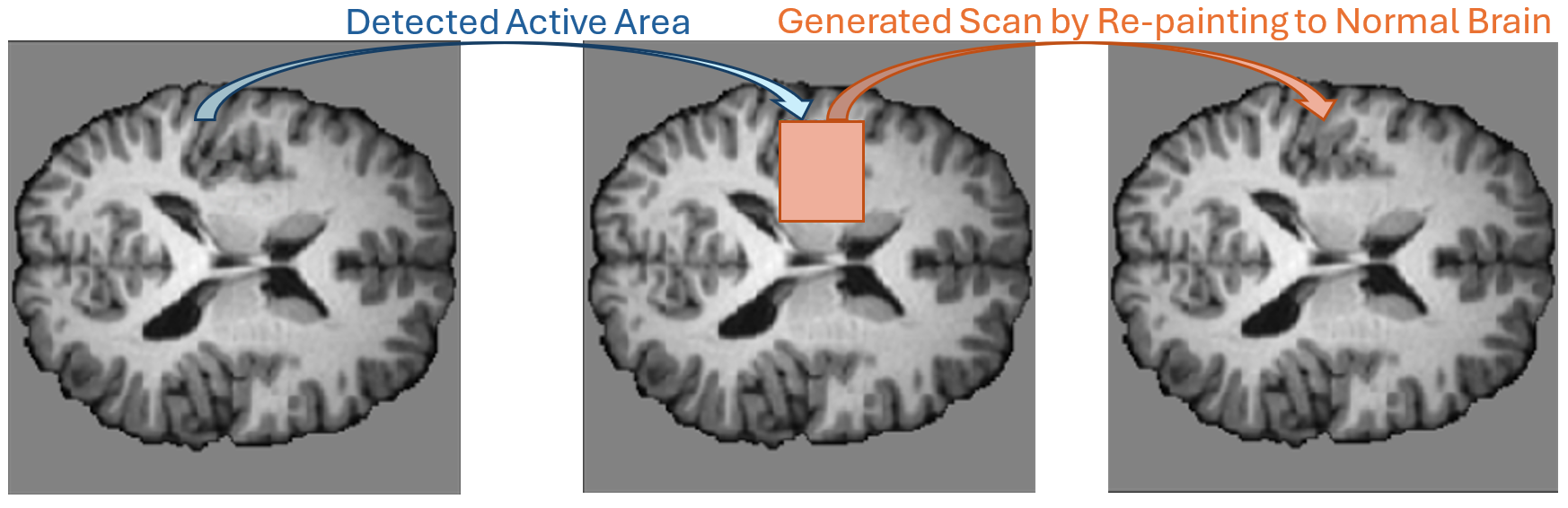
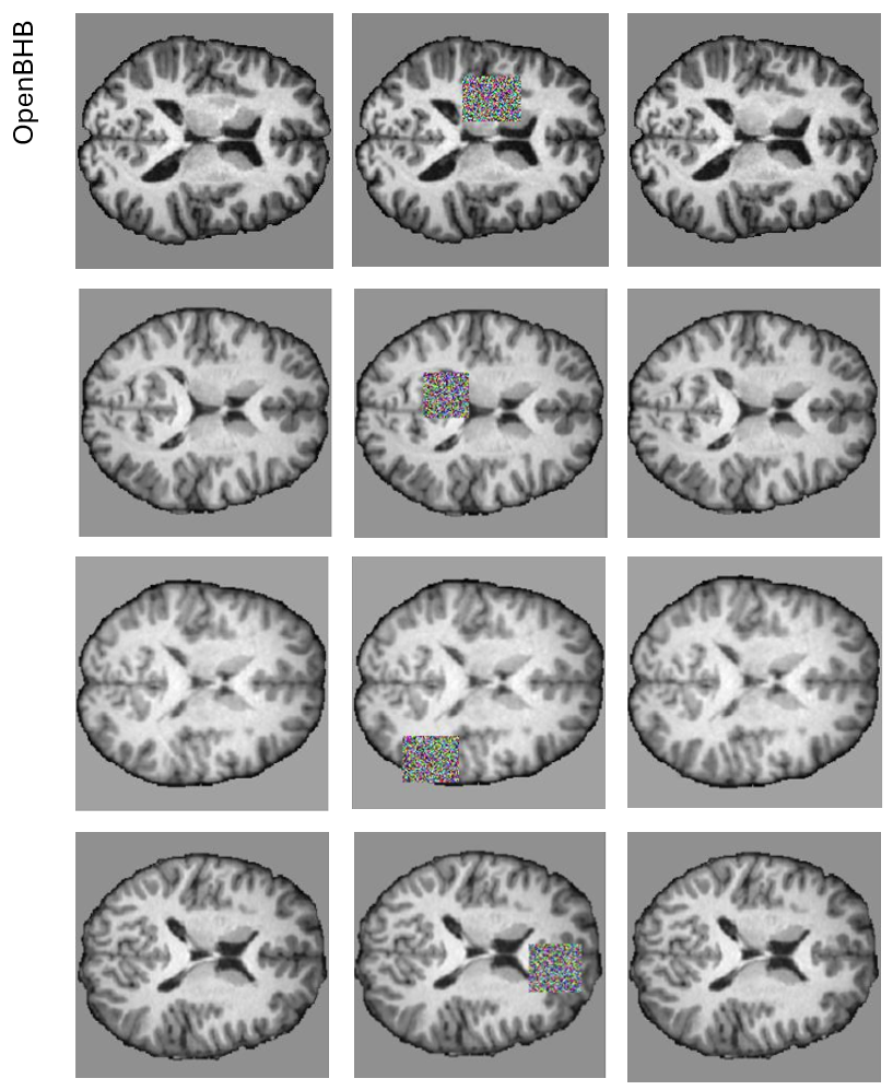
This project applies deep generative modelling to brain radiology scans to characterise and visualise how disease develops over time, supporting earlier and more interpretable assessment.
Output
- Rizia, M., Xu, C., Roberts, J., Barrett, L., Karunasena, S., Edelstein, S., & Suominen, H. Understanding the Development of Disease in Radiology Scans of the Brain through Deep Generative Modelling, IEEE BIBM 2024 — IEEE Xplore
Clinical collaborators:
- I-MED Radiology Network, Australia
- Royal Brisbane and Women’s Hospital, Brisbane, QLD, Australia
- Monash Health, Clayton, Victoria, Sydney, NSW, Australia
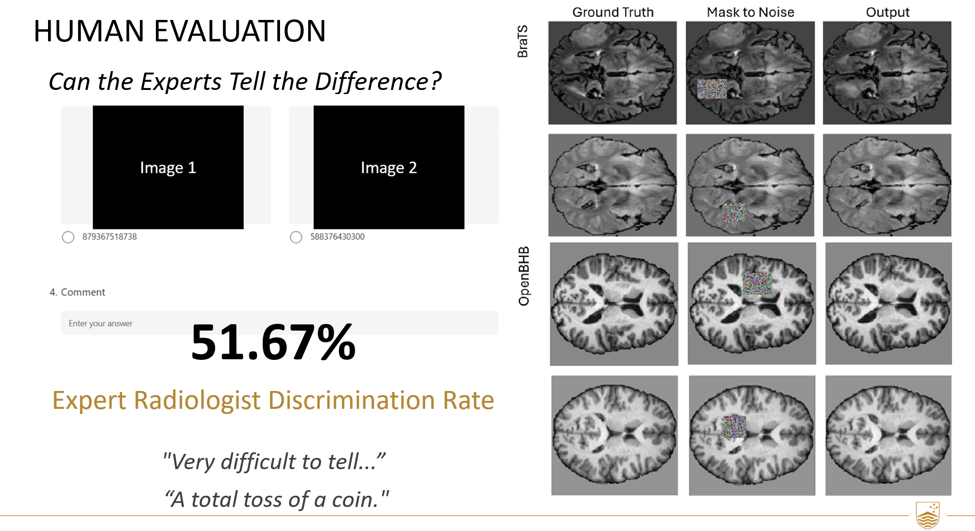
Dataset: BraTS & OpenBHB In a blind expert evaluation, radiologists could distinguish the generated scans from real MRI only 51.67% of the time — close to chance — indicating the synthesised scans were near-indistinguishable from genuine ones.